TL;DR
- What the FDA count actually measures, and what it does not, including the 76% radiology share and the 1,104 devices behind it. Jump to Where the Numbers Actually Stand.
- Three forces that made radiology the first specialty to run into an AI wave, from data availability to reimbursement precedent. Jump to Why Radiology Became the Center of the AI Debate.
- Why the replacement debate keeps missing the daily work of a radiologist, including the edge cases where clean input data does not exist. Jump to Why the AI Replace Radiologists Argument Keeps Missing the Actual Work.
- The AI use cases now clearing renewals in production, from worklist triage and medical image object detection to report drafting. Jump to What AI in Radiology Is Actually Doing Today.
- Four product opportunities that live around the model, and why the workflow is the larger surface area. Jump to Product Opportunities Beyond Detection.
- How vendor consolidation is reshaping ROI math for standalone AI purchases and what that means for single device vendors on the acquisition curve. Jump to Where the Vendor Market Is Concentrating.
- The questions hospital buyers actually ask during procurement, which run past sensitivity and AUC into workflow, liability, and reimbursement. Jump to What Buyers Are Actually Evaluating.
- Three shifts to watch over the next 24 months on foundation models, quality monitoring, and accreditation, plus the labor economics question underneath them. Jump to Where Machine Learning and Radiology Are Heading Next.
- The deployment path that keeps a first pilot alive to renewal, and the single test at the end that decides survival. Jump to How to Deploy AI in Radiology Without Adding Work.
- Seven decisions that separate the products that get renewed from the ones that get shelved after a pilot. Jump to What Teams Building for This Market Should Do.
Every few months, the same question comes back into circulation. Will AI replace radiologists? Search behavior around the phrase itself shows how much anxiety and curiosity now sits around the specialty.
Radiology draws this framing more than any other specialty for a specific reason. From the outside, the job looks like image inspection at scale, and image inspection reads to non-clinicians as an automatable task. Anyone who has worked inside a reading room knows how little of the actual job that appearance captures.
The question is dramatic. It is also the wrong first question for AI vendors, radiology groups, hospitals, and health tech teams trying to build useful products.
A more productive framing asks where artificial intelligence in medical imaging removes friction from the radiology workflow without removing clinical accountability. That framing produces a very different set of answers. It also produces a much more interesting market map.
From an AI transformation perspective, the opportunity is workflow redesign around trustworthy human and AI collaboration. Attaching a model to PACS and hoping adoption happens has produced many of the failed pilots in the market so far.
Where the Numbers Actually Stand
Every discussion of artificial intelligence in medical imaging now runs into the same headline number. Radiology holds 76% of every AI enabled medical device the FDA has ever cleared. As of the FDA’s most recent update covering authorizations through the end of 2025, that comes out to 1,104 radiology devices among 1,451 total authorizations. Growth has been steep. The list held 692 devices in October 2023 and crossed 1,500 in the first quarter of 2026.
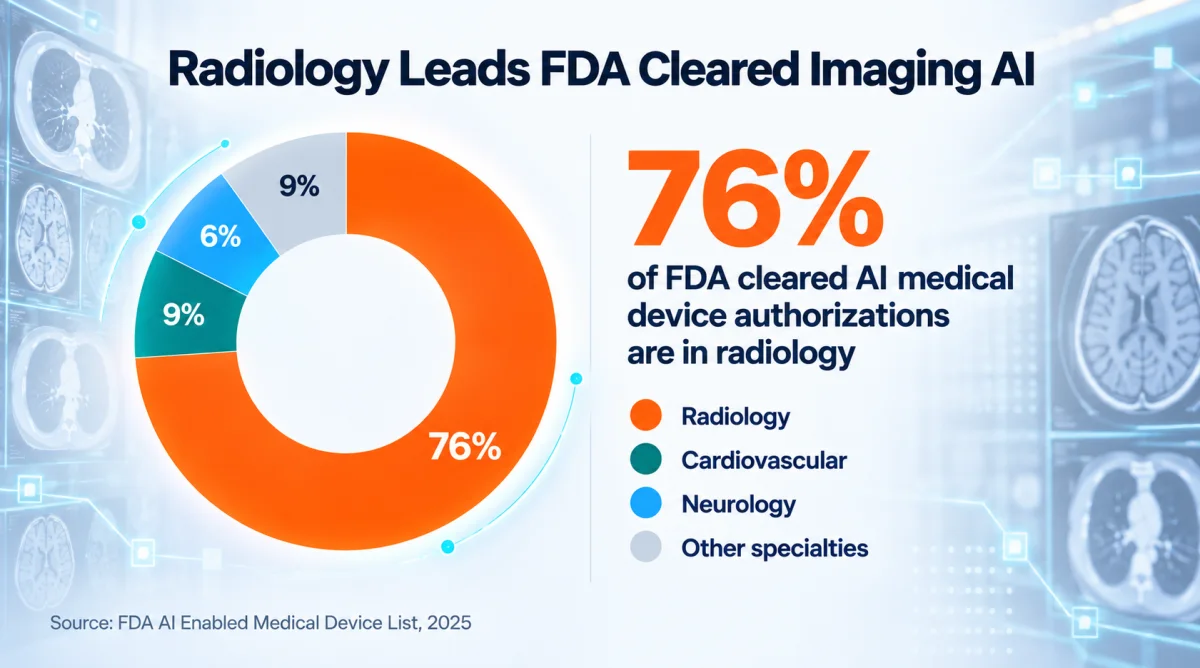
Fig 1 – FDA cleared AI medical device authorizations by specialty since 1995. Radiology holds 1,104 of 1,451 total. The top three specialties (radiology, cardiovascular, neurology) account for 90.6% of the market. Every other clinical specialty sits in a long tail.
The number gets deployed to argue two contradictory positions. One camp reads 76% as evidence that radiology is closest to full automation. The other camp reads it as evidence that the specialty is drowning in narrow tools that vendors ship faster than hospitals can validate.
Both interpretations miss what the number actually measures. FDA clearance describes what can be sold. Purchase, integration, use, monitoring, and renewal sit outside what it measures.
The gap between authorization and clinical impact is where the interesting work sits.
The ACR published its own read on the same data in August 2026. Nearly 90% of surveyed ACR members reported using some form of AI in practice. The Commission on Informatics developed the first practice parameter for imaging AI, adopted by ACR Council in May of 2026. Adoption is real. Governance is beginning to catch up. The question of what AI in medical imaging is actually doing has finally moved past the demo stage.
Why Radiology Became the Center of the AI Debate
Three forces put radiology at the front of the AI queue.
The first is data. CT, MRI, X-ray, ultrasound, mammography, PET, and fluoroscopy already live in digital infrastructure. Every study produces DICOM files with structured metadata, standardized viewing conventions, and decades of retrospective archives that model builders can train on. Few other specialties have that head start. A dermatology dataset requires a photographer. A cardiology dataset requires a device. A radiology dataset already exists.
The second is task shape. A pulmonary embolism, an intracranial bleed, a pneumothorax, a fracture, a lung nodule, a mass, a stenosis, and a measurement task can each be framed as detection, segmentation, or classification. That framing is what made medical image object detection one of the earliest AI use cases in healthcare and what continues to attract computer vision talent to the field. Detection is the entry point to a larger product opportunity, and vendors that start with medical image object detection often end up building or acquiring the workflow around the model.
The third is market structure. The FDA’s 510(k) pathway is well trodden for imaging devices, and radiology has thirty years of AI regulatory precedent going back to R2 Technology’s ImageChecker in 1998. Vendors know the reviewers. Reviewers know the vendors. Payors have started attaching reimbursement to specific AI enabled workflows. Viz.ai‘s large vessel occlusion product secured a New Technology Add on Payment from CMS. HeartFlow FFRCT holds an NTAP as well. That kind of financial infrastructure exists in almost no other specialty.
Exposure and arrival are different milestones. Radiology is where AI has landed hardest, and the specialty has yet to reach arrival.
Why the AI Replace Radiologists Argument Keeps Missing the Actual Work
Ten years have passed since Geoffrey Hinton predicted in 2016 that radiologists would be obsolete within five to ten years. The window has closed. Radiology residency match rates are at record highs. The 2025 match saw radiology fill at 99.8% with high applicant demand. Hiring is up across academic and community groups. The ai replace radiologists argument keeps returning because it has never engaged with what a radiologist actually does.
A scan supplies evidence. A bounding box marks a candidate finding. A report interprets those findings, and a care plan acts on them. Radiologists combine imaging findings with clinical context, prior studies, protocols, artifacts, anatomy variants, disease progression, surgical history, and the ordering clinician’s question. They decide what matters, what is incidental, what needs urgent escalation, what needs follow up, and what is likely noise.
That work is what many AI demos leave out and what many deployments run into. A model may perform well on a benchmark and then struggle when a hospital changes scanner protocols, updates software, shifts patient mix, or routes images from a new site. A model may accurately identify a finding and still create extra downstream work if it flags too many low value abnormalities. A tool may be technically strong and still fail if it surfaces outside the radiologist’s natural workflow.
Input quality is the other reality the replacement conversation skips. Scheduled outpatient studies with cooperative patients and standardized protocols produce the clean data most AI models train on. Emergency department imaging often does not. Studies arrive from altered, intoxicated, pediatric, nonverbal, or actively crashing patients where positioning, motion, and clinical context all deviate from the training distribution. A significant share of the clinical volume that matters most falls into this messier category, and vendors who have not designed for that reality tend to see performance drop during evaluation.
The serious conversation around machine learning and radiology has already moved past model accuracy. The current gates are validation on local data, monitoring for drift, human machine interaction, governance, bias detection, workflow fit, and clinical utility. The multi society statement from ACR, CAR, ESR, RANZCR, and RSNA lays out the same list in practice terms. Regulatory clearance is an entry ticket. Local success requires additional work in every one of those seven areas.
What AI in Radiology Is Actually Doing Today
The near term opportunity is disciplined deployment of AI into high friction points of the imaging lifecycle. Several use cases have moved from pilot to production over the past three years.
Worklist triage
Worklist triage for urgent findings has produced the clearest ROI story in the market so far. Suspected intracranial hemorrhage, pulmonary embolism, pneumothorax, and large vessel occlusion stroke are the anchor indications. Aidoc reports over 1,000 healthcare institutions using its triage suite. Viz.ai‘s LVO product secured CMS NTAP reimbursement based on published reductions in door to needle time for stroke patients. RapidAI operates in a similar category with cerebrovascular indications.
Medical image object detection
Nodules, fractures, bleeds, masses, calcifications, and device placement checks anchor the second high volume category. It is also the category most vulnerable to overcall and false positive fatigue when the vendor has not tuned the operating point for local prevalence. Fracture detection tools show pooled sensitivity around 90 to 92% and specificity around 91% across dozens of imaging studies. Chest imaging AI performance is more variable and depends heavily on the study population.
Segmentation and quantification
Segmentation and quantification for tumors, organs, vessels, cardiac structures, bone density, body composition, and disease progression has taken over hours of radiologist mensuration work in modern reporting workflows. Opportunistic screening on existing CTs for coronary calcium, hepatic steatosis, low bone density, and body composition markers is a growing category and one the ACR now flags as high impact.
Report drafting and impression generation
Report drafting and impression generation is the newest addition. Rad AI, Nuance DAX, and several health system pilots now generate draft impressions. The radiologist edits the draft. Writing every report from scratch is no longer the starting point. Vision language models and foundation models are starting to produce entire draft reports for radiologist review, and the ACR has flagged this as one of the most significant workflow shifts since PACS and voice recognition.
Operational infrastructure
Prior study comparison, change detection, protocol adherence quality control, and follow up tracking for incidental findings round out the operational use cases now in active deployment.
None of this requires the radiologist to disappear. All of it makes the radiologist more productive. The imaginative claim to build against is that radiology AI’s value extends past detection. It becomes an operating system for imaging departments, one that routes attention, compresses routine work, surfaces hidden risk, and turns every scan into training data for the next model.
Product Opportunities Beyond Detection
The most durable product opportunities in radiology AI live around the model. Detection is one moment in the workflow. The larger surface area is the workflow itself.
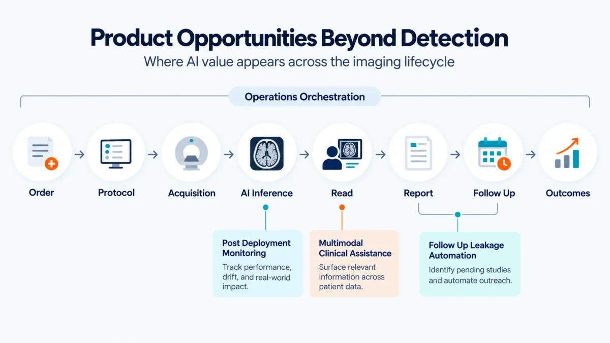
Fig 2 – Four product opportunities sit around the detection model. Each anchors at a different point in the imaging lifecycle, from order intake to outcome tracking.
Radiology operations orchestration
One opportunity is a radiology operations system that watches the full imaging lifecycle from order through outcome. That covers order intake, protocol assignment, acquisition quality, triage, read, report, communication, follow up, and outcome tracking. Most AI tools touch a single moment in that chain. The next category winner will orchestrate the whole chain and become the operational backbone for imaging groups.
Post deployment monitoring infrastructure
A second opportunity is post deployment monitoring infrastructure. Hospitals need to know whether a model still performs correctly after scanner changes, protocol updates, patient mix shifts, and software upgrades. Model drift dashboards, bias checks, and site specific performance reports are becoming necessary infrastructure for any practice running more than three AI tools in parallel.
Follow up leakage automation
A third opportunity is follow up leakage automation. Incidental findings are clinically important and operationally messy. Most findings get flagged in a report and then get lost in the handoff between radiology, referring physician, and patient. AI can identify findings that require follow up, route them to the accountable team, track completion, and close the loop. That is a compliance improvement, a revenue capture opportunity, and a patient safety product in one product.
Multimodal clinical assistance
A fourth opportunity is multimodal clinical assistance. The current generation of tools inspects images. The next generation will read the order, prior reports, labs, operative history, oncology notes, and comparison studies, and help the radiologist answer the actual clinical question the ordering physician asked.
The strategic lesson from this map is direct. Product durability follows workflow integration. A system that routes the right case to the right human with the right context at the right time is what earns renewal spend and long term expansion inside a healthcare buyer.
Where the Vendor Market Is Concentrating
Ownership of the radiology AI market is uneven. The top six vendors are shown below, counted from the FDA list through September 2025.
That is the concentrated end of the market.
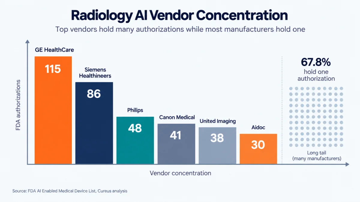
Fig 3 – Radiology AI vendor concentration. The top six vendors hold the bulk of authorizations. 67.8% of the 740 unique manufacturers on the FDA list hold only a single authorized device.
The long tail tells a different story. Cureus analysis of the full FDA list shows 67.8% of the 740 unique manufacturers have only a single authorized device. Just 13 companies account for 17.3% of all authorizations. The top three specialties, radiology, cardiovascular, and neurology, represent 90.6% of everything cleared.
Two implications matter for healthcare buyers.
First, the market is consolidating faster than the FDA count suggests. When a major imaging OEM ships a scanner, the AI ships with it. That changes the ROI calculation for standalone vendor purchases and makes multi vendor orchestration a real challenge for imaging IT teams.
Second, single device vendors are the most likely to be acquired or shut down within a three year window. Buyers evaluating a small vendor should assume that either the tool becomes part of a larger imaging OEM’s catalog or it disappears. Diligence questions should be built around that assumption from day one.
What Buyers Are Actually Evaluating
Many radiology AI vendors undersell the hardest part of the product. They lead with sensitivity, specificity, AUC, or benchmark performance. Those numbers matter. A hospital buyer is asking a different set of questions.
Hospital and imaging center evaluations now cover ground the pitch deck rarely reaches.
- Will this work on our scanners, protocols, patient population, and edge cases?
- How does it integrate with
PACS, RIS, EMR, dictation, and the existing worklist? - Can radiologists trust the output without becoming dependent on it?
- What happens when the model is wrong, and who owns the liability?
- Who monitors performance after deployment, and against what baseline?
- Can the vendor prove ROI without producing downstream noise like unnecessary follow up scans?
- Does deployment measurably reduce turnaround time, length of stay, missed findings, radiologist fatigue, or follow up leakage?
- Can compliance, IT, legal, radiology leadership, and clinicians all live with the workflow?
These are the questions Clixlogix would work through with a healthcare team before recommending a model, a vendor, or a custom build. Vendors who answer them with data close renewals. Vendors who reroute the conversation back to model metrics run pilots that die at contract time.
Reimbursement is where the ROI conversation gets real. Very few radiology AI tools carry independent Category I CPT codes. Most sit under Category III CPT codes that describe emerging technology and pay minimally if at all. The exceptions matter. Viz.ai secured NTAP reimbursement for its LVO stroke product. HeartFlow FFRCT holds an NTAP for coronary analysis. Almost everything else has to prove ROI through operational efficiency, throughput improvement, or downstream care benefits. That is a longer sale than a demo of the model detecting X.
The transformation work is where an AI implementation partner earns its fee. It includes mapping the clinical workflow, designing the human in the loop process, building the governance protocol, monitoring data drift, creating evaluation dashboards, training users, and turning AI output into operational improvement. The full transformation often involves integrating AI into legacy application environments that were not built with AI orchestration in mind.
In radiology the algorithm is one component. Trust at scale is the actual product.
Where Machine Learning and Radiology Are Heading Next
Three shifts are worth watching over the next 24 months.
Foundation models move to production
Foundation models and vision language models will move from research to production. Draft report generation is already piloting in academic centers. Vendors like Rad AI, Aidoc, and RadNet’s DeepHealth are shipping products that produce structured reports for radiologist review. The ACR is treating this as the most significant workflow change since PACS and voice recognition. The economics look different too. A vendor selling a report drafting tool is targeting radiologist time savings, which converts to hard ROI at any group where read volume is capacity constrained.
Quality monitoring becomes table stakes
Quality monitoring is becoming table stakes. The ACR Assess AI service, described in the August 2026 Commission on Informatics update and in the JACR, combines data from the radiologist’s report, DICOM header, and AI output to produce real world model performance metrics. Participating sites use those metrics to decide whether to deploy new models, keep existing ones, or take them offline. Any vendor selling into a large practice in 2027 should assume post market monitoring is part of the contract.
AI accreditation arrives
Accreditation is coming. ACR Council approved exploration of AI accreditation in 2026. Once that program launches, buyers will start asking whether a vendor’s tool has been evaluated inside the accreditation framework. That is a new gate that did not exist even two years ago and one that will separate serious deployments from opportunistic pilots.
The labor economics question underneath
Underneath these three shifts is a demographic reality. Imaging volume is growing faster than radiologist supply. ACR workforce data shows rising vacancy rates across community and academic practices, and imaging volume has continued to grow at roughly 4 to 5% annually in the US. Capacity pressure is the driver of AI adoption in radiology. Anything that credibly extends radiologist reach will accelerate under that pressure.
A second order question sits underneath the capacity math. If AI raises the productive read capacity of a radiologist by 20 to 30%, staffing models, group compensation structures, and training pipelines all move. Some of that gain gets absorbed by the existing capacity deficit. Some of it changes the economics of who does what work. That conversation is already underway inside larger practices and will surface publicly over the next few years.
How to Deploy AI in Radiology Without Adding Work
For teams running a first radiology AI pilot, model selection is the last decision to make. The right starting point is a workflow pain that everyone in the group already agrees is real.
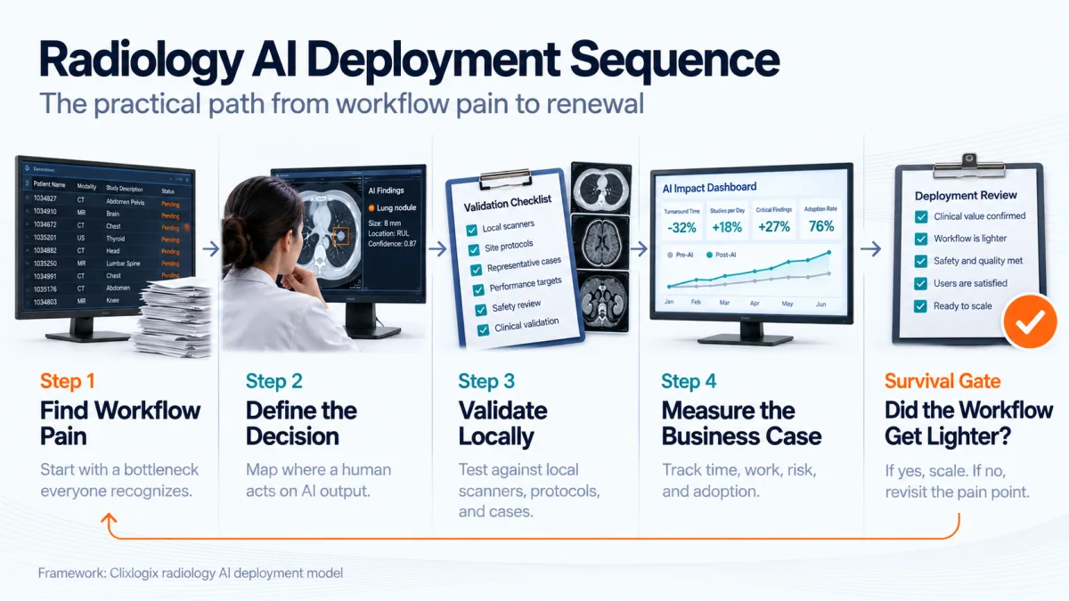
Fig 4 – The deployment sequence that keeps a first radiology AI pilot alive to renewal. Four build stages feed a single survival gate. Pilots that fail the gate loop back to workflow pain re-selection. Model retraining sits outside that loop.
1. Start with one high friction use case
Delayed urgent reads, missed follow up on incidental findings, inconsistent measurements, reporting burden, backlog on second reads, protocol errors, and lack of turnaround time visibility are all defensible starting points. Whichever one the group agrees on, the pilot has a clean success definition from day one.
2. Define the human decision point
Who sees the AI output? When do they see it? What can they do with it? Can they override it? Is the override documented? Does the output create a task, a report suggestion, an alert, or a background prioritization? None of these questions have universal answers, and getting them wrong is what turns a good model into an ignored tool.
3. Test on local retrospective data before going live
A tool that performs well in published studies may behave differently on local scanners, local protocols, local disease prevalence, and local reporting habits. The first 500 studies at any site are what makes or breaks clinical trust in the tool.
4. Measure the business case in operational terms
Turnaround time, read volume, length of stay, follow up completion, radiologist satisfaction, alert burden, and downstream testing are the metrics that hold up at renewal. Model accuracy metrics are a supporting measure.
The survival test
The single test for pilot survival at renewal is whether the workflow got lighter. That is the practical test.
What Teams Building for This Market Should Do
For teams building or buying artificial intelligence in medical imaging, the entry bar is now higher than a working prototype. Seven decisions separate the products that get renewed from the ones that get shelved after a pilot.
- Define the clinical problem with radiologists before defining the model. The best vendors spend enough time inside a partner site to understand workflow, exceptions, incentives, and failure modes before shipping.
- Validate performance on local, messy, representative data. Benchmark AUC is a starting point. Local operating point tuning is where clinical utility gets made.
- Integrate where decisions already happen. Any product that requires a second monitor, a second login, or a second workflow starts at a permanent adoption disadvantage.
- Design for override, uncertainty, escalation, and audit. Radiologists are the accountable party. The tool needs to make it easy for them to disagree with it.
- Monitor model performance over time, including drift, scanner specific issues, and demographic bias.
ACR Assess AI is one of the leading emerging reference points. - Measure business and clinical outcomes alongside model metrics. Turnaround time, length of stay, missed findings, follow up completion, and radiologist read volume are the metrics that hold up in a renewal conversation.
- Treat trust as a product feature. Explainability, provenance, and clear failure modes belong in the roadmap the same way core detection accuracy does.
The next generation of radiology AI will focus on reliable operations. It will help radiologists practice at the top of their license. It will help hospitals scale expertise at a moment when imaging volume is growing faster than radiologist supply. It will help vendors build businesses that survive past the pilot budget line.
Machine learning and radiology already belong together. The remaining question is whether the systems being built are imaginative enough to improve medicine and disciplined enough to earn medicine’s trust.
Work With Clixlogix
Clixlogix works with healthcare teams on AI strategy, feasibility validation, clinical workflow design, and production AI system development for imaging and adjacent clinical operations. Our engagements cover the full lifecycle from opportunity identification through deployment, monitoring, and iteration. If you are scoping an AI initiative and want an implementation partner who thinks about workflow, monitoring, and ROI from the start, contact us for an initial conversation.
Get in Touch