TL;DR
The Cobb angle project started as a measurement problem. A medical imaging software company had a scoliosis workflow that was slowing radiologists and frustrating referring orthopedic surgeons. Manual Cobb angle measurement added several minutes per study. Free text dictation had to be reshaped into a 14 field scoliosis template. Prior report comparison required manual PACS search.
Every one of those bottlenecks sat in the workflow around the measurement.
Clixlogix built the AI module inside the client’s existing desktop recorder and PACS workflow. The radiologist opened the study the same way. The AI module retrieved the study from PACS through DICOM query and retrieve. It returned accepted measurements to PACS as DICOM Structured Report objects. It returned visual measurement lines and vertebral labels as GSPS overlays. Study orders arrived as HL7 ORM messages. Signed results left the reporting stack over HL7 ORU carrying structured report fields and the DICOM SR reference.
That build reached 1.8 degrees mean absolute error against consensus radiologist reads. 91% of measurements landed within 5 degrees. Report turnaround moved from around 36 hours to under 10 hours. Template field completeness rose from 62% to above 90%. The non functional requirement set the path from study open to populated sidebar under 10 seconds.
That is the integration shape this article explains.
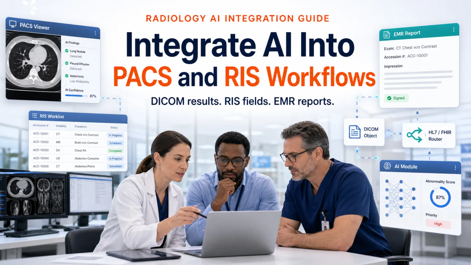
This is written for healthtech product owners and radiology IT teams deciding where AI output should live. If you are building an AI imaging feature, understand where artificial intelligence in medical imaging actually stands first, then map the systems (PACS, RIS, EMR), the standards (DICOM, HL7, FHIR), and the reader surface (viewer behavior, latency, sign off) before production.
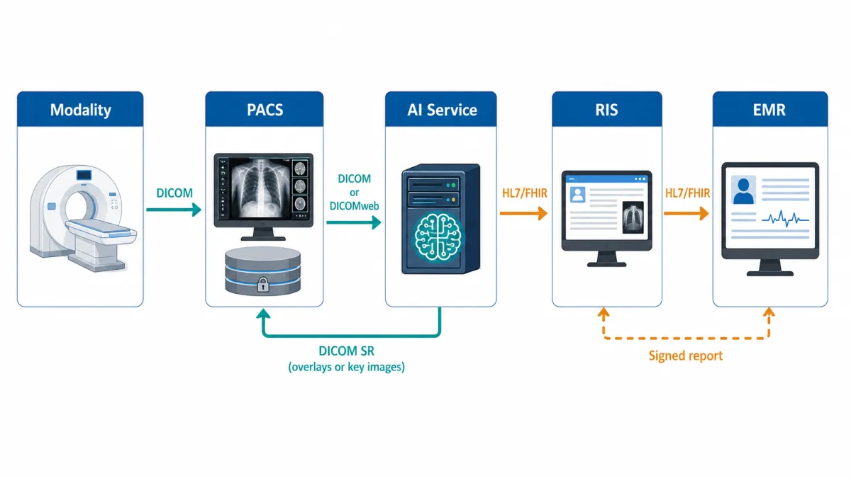
Fig 1 – A practical AI imaging flow. PACS holds the study and DICOM result objects. RIS and the reporting stack handle structured fields and report status. EMR receives the signed result.
The architecture in plain terms
An AI imaging tool needs three routes.
First, it needs a study intake route through DICOM routing, DICOM query and retrieve, DICOMweb, or viewer context. Second, it needs a result return route. Image derived output should go back as a DICOM object when the result needs to travel with the study. Third, it needs a clinical result route. RIS needs structured fields when AI affects report content, worklist state, QA routing, or operational analytics. The EMR receives the signed clinical result through the site’s normal result interface.
The Royal College of Radiologists makes this point directly in its guidance on integrating AI with RIS and PACS workflows. AI findings should be communicated to PACS through DICOM and to RIS through HL7. The guidance also says AI analysis should be complete and available on PACS before the human reporter starts interpretation.
RSNA’s Radiology Reimagined program follows the same direction. It shows AI in realistic radiology scenarios using standards such as IHE AI Results, IHE AI Workflow for Imaging, interactive reporting, common data elements, and FHIRcast. Buyers want a result that moves through the same clinical systems that already move the study and report.
Native or bolt on integration
Buyers usually frame the first decision as native or bolt on.
Native integration puts the AI result inside the PACS viewer the radiologist already uses. Findings appear in the reading surface next to the images, with no second application to open. Sites favour this because it protects the read and keeps training effort low. It also ties your product to specific viewer vendors and their release cycles, so each new site can mean new work.
Bolt on integration runs the AI service alongside PACS and returns results through standards based routes. The radiologist sees results as DICOM objects in the viewer, as structured fields in the report, or in a separate review surface. This travels across sites more easily because it depends on DICOM and HL7, which every imaging site already runs.
The four intake patterns in the next section are the practical expression of that choice. Viewer context integration sits at the native end. DICOM routing, query and retrieve, and DICOMweb sit at the bolt on end.
Larger sites often add a third layer. AI orchestration platforms such as Blackford, Aidoc, deepc, CARPL, and the Nuance AI Marketplace sit between PACS and a catalogue of AI applications. The platform handles study matching, routing rules, result write back, and monitoring for every algorithm the site runs, so the site integrates once instead of once per vendor.
For a single AI feature, direct integration usually ships faster. For a site already running several algorithms from different vendors, the orchestration layer removes repeated integration work and gives governance one place to live. Ask early which model your buyer runs. A site standardised on a marketplace will expect your product to arrive through it, and that changes your packaging, your result format, and your commercial route.
Where AI receives the study
You have four common intake patterns. Pick the one that matches when the result is needed.
| Placement pattern | How the AI receives the study | Best fit | What to test |
|---|
| DICOM routing after acquisition | PACS or modality routes matching studies to the AI service | Triage, quality checks, batch measurements, segmentation | Routing rules, duplicate sends, study type filtering, failed retries |
| DICOM query and retrieve | AI service retrieves the study from PACS when worklist or viewer context opens | Reader assist, prior aware tools, on demand measurements | Study open latency, accession matching, missing prior behavior |
| DICOMweb | AI service uses QIDO-RS, WADO-RS, or STOW-RS where supported | Cloud archives, modern viewers, web based imaging products | Site support, auth model, object retrieval speed, fallback path |
| Viewer context integration | PACS viewer or recorder passes the active study context | Sidebar assistance during interpretation | Viewer compatibility, context sync, timeout handling, user identity |
DICOM routing works well when the AI result can be prepared before the radiologist opens the study. DICOM query and retrieve fits tools that react to the active read. DICOMweb fits newer archives and web viewers. The DICOM PS3.18 Web Services standard defines web service access patterns for DICOM resources, including query, retrieval, and storage. Viewer integration fits products that assist the reader inside the workstation. Keep that integration boundary small. Pass study identifiers, user context, and result panel state. Keep the clinical result in the standards based flow.
What AI writes back to PACS
PACS is the imaging archive. If the AI output comes from the image and needs to remain connected to the study, PACS should usually receive a DICOM result object.
The current DICOM Standard is broad. For AI imaging products, the practical question is which DICOM object carries which result.
| AI output | PACS object route | Use when | Buyer concern |
|---|
| Numeric measurement | DICOM Structured Report | The result needs structured storage and image references | Does the receiving viewer display or expose it cleanly |
| Visual line, label, or annotation | GSPS | The viewer needs to show overlays tied to the source image | Does every target viewer render it as expected |
| Segmentation | DICOM SEG | The output is a mask, contour, organ, lesion, or region | Does the downstream viewer support the segmentation object |
| Review image | Key image or secondary capture | Human review needs a visible summary | Is the result still machine usable elsewhere |
| Internal confidence or QA flag | AI platform log and RIS field where relevant | Product monitoring, QA routing, or audit | Is the flag governed and traceable |
DICOM Structured Reporting is a strong fit for measurements because it can carry coded content, references to source images, and structured values. The DICOM information object definitions include the object model that SR and presentation states rely on. GSPS is useful when the result needs to be seen as an overlay. In the Cobb build, endplate lines and vertebral labels returned as GSPS overlays bound to the source study.
IHE’s AI Results profile points to the same operational need. AI results need to be stored, retrieved, and displayed in the radiologist’s reading environment.
What returns to RIS
RIS owns the operational radiology workflow. It tracks the order, accession, scheduling, modality context, report state, reader assignment, QA flags, and result lifecycle. If the AI result changes how the case moves, RIS needs to know.
Use RIS or the reporting stack for structured report fields, triage state, QA flags, acquisition quality warnings, report completeness checks, billing support fields, and result status events.
HL7 v2 remains common here. The HL7 v2.5.1 Observation Reporting chapter describes ORU messages for transmitting observations and results of diagnostic studies. Orders and order context often arrive through HL7 order messages, including ORM in older deployments.
FHIR may appear around newer EMR integration, patient facing products, and API based workflows. The FHIR ImagingStudy resource represents information about a DICOM imaging study. The FHIR DiagnosticReport resource represents the report. The FHIR Observation resource can represent measurements and simple assertions.
Keep the distinction clear. DICOM carries the image evidence. RIS carries operational report workflow. FHIR can expose approved clinical data through an EMR friendly API where the site supports it.
Do this mapping before interface build. It prevents late rework.
| System | What belongs there | Why it belongs there | Example |
|---|
| PACS or VNA | Studies, image derived objects, DICOM SR, GSPS, SEG, key images | The output stays attached to the source image | Cobb angle SR plus GSPS overlay |
| RIS | Orders, accession state, worklists, report workflow, structured fields, QA flags | Radiology operations stay consistent | Cobb angle field pre populates a scoliosis template |
| EMR | Signed clinical report, approved observations, report references | Care team reads the final clinical record | Signed report delivered through HL7 ORU or mapped FHIR resources |
| AI platform | Model metadata, confidence, inference logs, drift signals, override feedback | Product team monitors and improves the model | Override queue for retraining review |
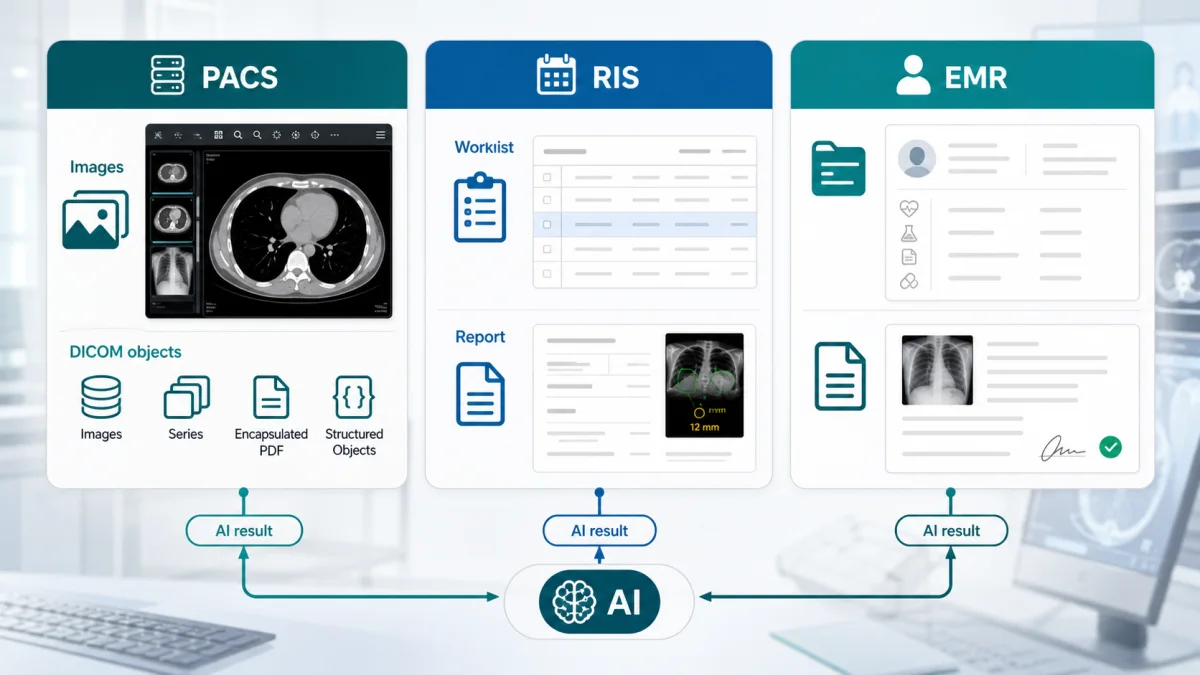
Fig 2 – PACS, RIS, EMR, and the AI platform should each own a different part of the workflow.
The Cobb build as a worked example
The Clixlogix Cobb angle case study gives a concrete version of this architecture.
The client operated an AI based medical imaging platform serving radiology groups and orthopedic specialist clinics. The scoliosis module had to detect end vertebrae, propose landmarks, compute Cobb angles, support S curves, handle post surgical hardware, and work across standing spine radiographs and EOS studies. The radiologist had to review every measurement. The signed value had to remain the radiologist’s value.
Here is the actual integration path:
| Workflow step | Integration choice | Why it mattered |
|---|
| Study intake | DICOM query and retrieve against PACS | The AI service received the study without changing the radiologist’s worklist |
| AI result to PACS | DICOM SR | Measurements, vertebrae, confidence scores, and model version stayed attached to the study |
| Visual explanation | GSPS overlays | Endplate lines and vertebral labels remained visible in supported DICOM viewers |
| Orders | HL7 ORM | Study order context drove template selection |
| Signed results | HL7 ORU | Structured report fields and the DICOM SR reference left the reporting stack |
| Reader workflow | Desktop recorder and PACS sidebar | The radiologist reviewed, accepted, adjusted, or overrode inside the existing workflow |
| Governance | Audit log and model version traceability | Every measurement and override remained traceable |
Validation ran before production on 2,400 studies. The corpus covered pediatric and adult standing spine radiographs, EOS studies, Risser stage, curve severity, acquisition modality, and post surgical status. The reference standard was consensus review by two board certified radiologists, with orthopedic spine surgeon adjudication when disagreement exceeded 3 degrees.
The production release cleared the agreed clinical thresholds:
| Metric | Result |
|---|
| Mean absolute error against consensus | 1.8 degrees overall |
| Measurements within 5 degrees of consensus | 91% |
| Pediatric slice mean absolute error | 2.4 degrees |
| Adult slice mean absolute error | 1.6 degrees |
| EOS slice mean absolute error | 2.0 degrees |
| S curve identification accuracy | 94% |
| Post surgical hardware detection recall | 97% |
| Study open to populated sidebar requirement | Under 10 seconds |
| Report turnaround | Around 36 hours to under 10 hours |
| Template field completeness | 62% to above 90% |
| Referring surgeon callbacks for missing fields | Over 70% reduction |
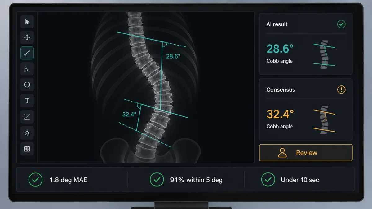
Fig 3 – The Cobb build combined workflow integration with measurable clinical and operational results.
The key lesson is simple. The result stayed in the systems the site already trusted. PACS held the imaging evidence. The reporting stack received structured fields before sign off. HL7 carried signed results out. The AI platform kept the model metadata, confidence scores, overrides, and retraining feedback.
Failure modes to plan for
Most AI imaging pilots fail in ordinary places such as routing, worklists, latency, viewer display, and signed value traceability. Test these risks early.
| Failure mode | What it looks like | How to reduce it |
|---|
| Wrong study routed | AI receives non target protocols or misses eligible studies | Test routing rules against real study descriptions and modality metadata |
| Accession mismatch | Result cannot attach cleanly to the order or report | Reconcile accession number, study UID, patient ID, and order ID in shadow mode |
| Worklist disruption | AI creates unexpected status changes or duplicate events | Keep worklist state changes explicit and test cancellations, addenda, and corrected reports |
| Viewer display gap | SR, GSPS, SEG, or key image output fails to render consistently | Test every target viewer and define fallback display behavior |
| Latency at study open | Sidebar result appears after dictation has started | Pre compute where possible and set strict on demand timeouts |
| Proprietary result path | Result lives only in the AI dashboard | Return clinical outputs through DICOM, HL7, or FHIR paths the site governs |
| Governance gap | Model version, confidence, and override history are hard to reconstruct | Store version, input references, user action, final value, and timestamp |
RCR guidance calls out the same risk at the workflow level. AI should integrate into existing RIS and PACS workflows in a way that avoids extra burden on radiologists. RSNA’s interoperability coverage makes the vendor case too.
Shadow testing before clinical release
Shadow testing is where you find workflow defects before clinicians rely on the result.
In shadow mode, the AI service receives real workflow traffic or a representative replay. Output goes to a test destination, QA queue, or internal dashboard. Your team measures routing, identity matching, mapping, latency, viewer behavior, clinical agreement, and failure handling.
Use this table as the launch gate.
| Shadow test area | What you measure | Release signal |
|---|
| Routing coverage | Studies sent, skipped, duplicated, misclassified | Eligible studies route consistently |
| Identity match | Patient ID, accession, study UID, order, report context | Result attaches to the correct case |
| Result mapping | DICOM, RIS, HL7, FHIR, report fields | Fields land where the site expects them |
| Viewer behavior | SR, GSPS, SEG, key image display | Target viewers show the result clearly |
| Workstation latency | Time from study open to useful result | Reader assist meets the agreed budget |
| Clinical validation | Agreement against consensus or signed reports | Performance holds across clinical slices |
| Failure behavior | Timeout, low confidence, missing priors, failed write back | Users see safe and predictable states |
| Governance | Model version, audit log, override capture | QA and regulatory review can reconstruct the case |
Radiology AI guidance stresses implementation and monitoring as part of responsible clinical use. The ACR Data Science Institute also frames AI around use case definition, validation data, clinical implementation, monitoring, and regulatory awareness.
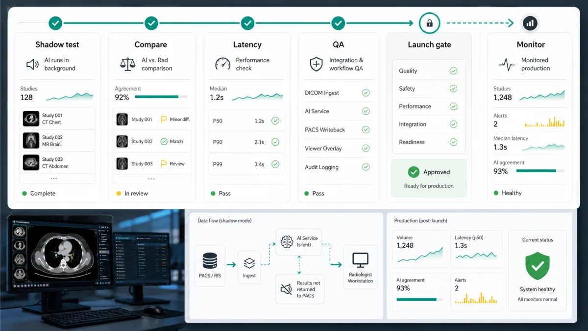
Fig 4 – Shadow testing should prove routing, mapping, latency, viewer behavior, clinical agreement, and governance before clinical release.
Standards checklist for product owners
Ask these questions before build.
| Question | Why it matters |
|---|
| Which DICOM path retrieves the study | Determines routing, latency, auth, and site compatibility |
| Which DICOM object carries the result | Determines viewer behavior and downstream reuse |
| Which RIS field receives structured output | Determines report template behavior and worklist safety |
| Which HL7 messages carry orders and results | Determines how the feature fits live hospital interfaces |
| Which FHIR resources apply | Determines API use for EMR, data lake, or patient facing workflows |
| Which identifiers are authoritative | Prevents accession, study UID, and patient matching faults |
| Which user signs the value | Keeps clinical responsibility clear |
| Which audit events are retained | Supports QA, support, validation, and regulatory review |
| Which failure states are visible | Prevents silent misses and unsafe assumptions |
The FDA Clinical Decision Support Software guidance is also relevant when your feature affects clinical decisions. FDA’s digital health materials explain that software analyzing medical images may fall inside device oversight depending on intended use. Bring regulatory review into the workflow discussion early.
How to decide where each result lives
If the result is a measurement tied to a source image, plan a DICOM SR path. If the result is an overlay or label, plan GSPS. If the result is a segmentation, plan DICOM SEG. If the result is a triage state, decide whether it changes the RIS worklist, the reporting queue, or a QA dashboard. If the result is final clinical content, route it through sign off.
Some AI outputs are invisible infrastructure. A study quality check can route to a technologist queue. A prior report retrieval signal can help the reporting stack. A measurement proposal belongs in front of the radiologist. A signed observation belongs in the final result path.
Triage has value before the read. Reader assist has value during the read. QA has value after the read. Analytics has value after aggregation. The timing determines whether you should run inference after acquisition, at worklist open, at viewer open, or after sign off.
| Result timing | Typical user | Best route | Design priority |
|---|
| Before read | Radiologist, coordinator | DICOM routing plus RIS worklist flag | Completeness and priority accuracy |
| During read | Radiologist | PACS viewer, sidebar, report template | Latency and clear review controls |
| At sign off | Radiologist, reporting stack | RIS and HL7 result path | Structured field mapping |
| After sign off | QA, governance, product team | AI platform and analytics layer | Monitoring and drift detection |
| For EMR consumption | Referring clinician, care team | Signed report, HL7 ORU, FHIR resources where supported | Approved clinical context |
How Clixlogix approaches AI imaging integration
Clixlogix builds healthcare software around the systems buyers already run. For imaging AI, that means DICOM objects, PACS viewer behavior, RIS worklists, HL7 order and result flows, FHIR paths where appropriate, PHI boundaries, validation evidence, and audit trails. Our AI software development team maps model output to workflow, standards, and release gates.
Relevant healthcare builds include the AI Cobb angle measurement platform, a public health data platform, a cardiovascular AI care companion, and a HIPAA compliant healthcare portal.
The first workshop is usually a workflow map. We identify the study source, the system of record, the reader surface, the result destination, the sign off point, and the failure states. Then we map the AI output to the right standards path.
That work protects the product later. Your buyer wants to know how the feature handles old PACS profiles, mixed viewers, patient merges, corrected reports, low confidence cases, addenda, site specific templates, and model updates.
For an AI imaging feature, start with one question. Where will the signed result live, and what workflow has to trust it.
Mapping an AI imaging feature to a real clinical workflow?
We scope imaging AI as an integration problem first, covering DICOM objects, PACS viewer behaviour, RIS worklists, HL7 result paths, and the sign off point before any model work starts. If you are planning an imaging feature, talk to our healthcare team and we will map the workflow with you.